Ct scan protocol books pdf
Elbow CT: Positioning We don’t scan patients with their elbow down at their side Excess radiation across torso X-ray scatter decreases res. NEVER scan patients with their elbow laying on their abdomen Elbow moves with patient breathing Makes it impossible to get good reformats Instead get terrible images like this – WRONG WAY CT Scout . Anatomy Technical FOPH BBFF Monteggia Galeazzi …
High resolution CT is a scanning protocol in which thin sections (usually 0.625 to 1.25 mm) are acquired and reconstructed using a sharp algorithm (e.g. bone algorithm).
Scan Protocols chapter 1 CT Scan Protocol Chapter 1 CT Scan Protocols PROPHECY® Alignment Guides are patient specific instruments designed to improve total knee replacement results. One significant requirement for a successful case is adhering to the CT scan protocol. Engineers at Wright Medical Technology have determined the necessary scanning parameters which are described in …
Developing Dose and Protocol Monitoring from Scratch Ingrid Reiser, PhD DABR Department of Radiology The University of Chicago. Outline • CT protocol monitoring – Let’s build a protocol book! • CT dose monitoring. From scratch: status as of ~2015 • Fleet of 8-9 Philips CT scanners, mix of inpatient, outpatient, dedicated pediatric • Radimetricsinstalled, about 60% of CT studies
Szczykutowicz, Timothy P., et al. “Compliance with AAPM Practice Guideline 1. a: CT Protocol Management and Revie w —from the perspective of a university hospital.” Journal of Applied Clinical Medical Physics 16.2 (2015).
protocol uses a split bolus between the CT scan of the chest, abdomen, pelvis (120 mL Optira y 320), and CT of the carotids and Circl e of Willis (80 mL Optira y 320).
This CT scan protocol has been designed to provide scan centers’ personnel with easy-to-use instructions to obtain correct images with optimal quality that will be further used in the process of surgical planning and design of patient specific surgical instruments and implants
A CT (computed tomography) scan, also known as a “CAT” scan, uses x-rays to form pictures of the inside of the body. How is CT different from a regular x-ray? During a regular x-ray procedure, a stationary machine sends x-rays through the body to make a single “shadow”
T he combination of positron emis-sion tomographic (PET) scanners and computed tomographic (CT) scanners, or PET/CT scanners, provides coregistered images of anatomic and
The following cardiovascular CT scanning and injection protocols have been developed with the goal to derive theoretically sound and at the same time practicable scanning and injections protocols for the Siemens Sensation 64 multi-slice scanner at Stanford.
CT scan protocol for head and neck bone tissue Export Data www.xilloc.com The CT scan quality is critical for the creation and production of accurate patient specific implants, surgical guides and/or anatomical models. So please read and apply following steps with care to assure an optimal image quality. If there are any questions please do not hesitate to contact us at all times at +31 644
Computed tomography (CT) is the most commonly used cross sectional imaging tool. This is a starting page for some general articles about CT: evolution of CT scanners CT protocol standardized reports review areas computed tomography (physics…
For any given CT examination, the protocol will vary according the individual requirement of the imaging site. Factors that infl uence a specifi c site’s protocol parameters include the type of equipment available (e.g., 64-slice versus 16-slice), the setting (e.g., inpatient versus outpatient), and the particular preferences of the radiologists in charge. Like all fi elds of medicine, CT is
Caryn Damits, RT (R) CT POSITIONING AND LANDMARKS WHY IS THIS IMPORTANT? 1. Dose 2. Isocenter 3. Consistency with Stanford CT Scans Isocenter • In imaging physics and radiation oncology, the `isocenter` is the point in space through which the
netics and patient, contrast medium, and CT scanning factors associated with contrast enhancement and scan timing are presented and discussed. Published data from clinical studies of contrast medium and physiology are re- viewed and interpreted. Computer simulation data are analyzed to provide an in-depth analysis of various factors associated with contrast enhancement and scan timing. On the
Standard Imaging Protocols This document includes typical imaging protocols for diagnostic imaging. vRad teleradiologists expect to see imaging resulting from the use of these or substantially similar protocols in
.AAPM Protocol Document Importance of tailoring abdomen-pelvis CT protocols to size can not be over emphasized. GE User (n=1): Routine abdomen/pelvis CT protocol . 5 sec Detector configuration 16 x 1.375 kV 120 120 Noise Index & Img Thickn 11.25 mm Pitch 1.
CT BODY PROTOCOLS Standard CTA/CTV Multiphase Abdomen/Pelvis CTA Abdomen/Pelvis Adrenal Kidney Stone CTA Abdomen/Pelvis w/ Delay Pancreas Chest/Abdomen/Pelvis CTA Lower Extremity Runoff Pancreas & Pelvis Chest/Abdomen/Pelvis & Neck CTA Upper Extremity Runoff Pancreas & Chest/Abdomen/Pelvis Chest & Neck Liver 2 Phase Pelvis CTA Thorax/Abdomen Pelvis Liver 2 Phase & Pelvis CT …
MEDICAL PHYSICS Whole-Body PET/CT Scanning

BRINK CCTA Protocols Contrast Media Optimization
Generic monitoring of scan protocols in CT • monitoring of scan protocols is important – to detect unauthorized modifications – to detect accidental upgrades by service personnel – to make sure that optimization efforts are used in the field • detection of modified scan parameters could also be done retrospectively using continuous patient dose monitoring of your CT system
We believe that CT scanning is an effective and essential tools in treatment planning, basic understanding of physiology, and and tackling the ever-increasing …
The book is designed for the new CT technologist. Everybody who cross-trains into CT will, at Everybody who cross-trains into CT will, at some point in time, find this information useful.
CT Scan: Capturing the CT When a scan appliance will be used, two scans are required, one of the patient with the appliance seated and one of the scan appliance alone.
CT Protocols (PDF – The Table of Contents is linked to the corresponding pages for quick access) MRI Protocols (PDF – The Table of Contents is linked to the corresponding pages for quick access)
MANUAL OF PROCEDURES PART B CT TECHNICAL PROCEDURES. American College of Radiology Imaging Network CQIE Manual of Procedures Version 3.2 March 2013 Part B: 1 of 7 PART B CT TECHNICAL PROCEDURES 1. CQIE CT QUALIFICATION 1.1 Introduction The purpose of this chapter is to provide detailed information regarding the CT phantom scanning and quality control activities …
Whenever I give a lecture or presentation the most commonly asked question is “What’s your protocol?” To help answer those questions we have put together “our” protocols as well as adding CT Scan protocols from other institutions that we thought might prove useful.
Cardiac CT Angiography • Intravenous Contrast Material •Flow Rate •Concentration •Type •Duration (Volume) •Scan Delay
This book provides structured up-to-date information on all routine protocols used for multislice (multidetector row) CT. The volume contains a detailed technical section and covers the prevailing investigations of the brain, neck, lungs and chest, abdomen with parenchymal organs and gastrointestinal tract, the musculoskeletal system and CTA as
Overall this is an excellent little reference book to take along in clinical rounds.It will fit into your lab coat with no problems. Like it states it is intended for the Radiology Resident or the C.T. Technologist with some or little background on the basic principles.
Offering radiology services and exams: MRI, CT, X-ray, Open MRI, PET/CT, Nuclear Medicine, Ultrasound, Interventional Radiology, and Neurointerventional Radiology. Our sub-specialized radiologists offer teleradiology and professional read services for various medical groups and hospitals.
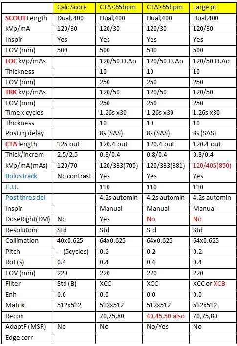
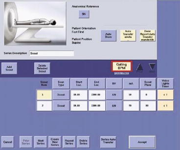
thoracic CT aortogram Gated scan, timing bolus Imaging protocol: prospective ECG-triggered CT aortogram ECG gated, prolonged scan duration AP and lateral scouts Right arm IV placement • Avoid streak artifacts across arch vessels Timing bolus below carina. ROI in descending thoracic aorta. PT sequential axial acquisition at timing bolus peak + 4 sec Contrast • Biphasic size based bolus
20/07/2010 · Children are more sensitive to radiation than adults. Computerized tomography (CT) consists of 25 % of all medical imaging. It was estimated that more than 2% of all carcinomas in the USA are due to CT scans. There is an ongoing focus on the reduction of CT scan radiation dose. Awareness about risk
•A radiologist protocols a scan •An institution has a master protocol book •The scanner comes with built in protocols •The patient was scanned with this protocol AAPM 2017 Computed Tomography Imaging: CT Protocol Management 4 We Need Definitions and Categorizations to Communicate Effectively Protocol – what’s in a name •DICOM Supplement 121: CT Protocol Storage •Published …
Imaging Location: Record CT scan – a specific protocol must be followed for each planning software. iDent.Page 4 Scan Appliance 4 P a g e Scan Appliance Technique ‐ Indications & Protocols: When is a Scan Appliance required? ‐Reconstructive cases ‐ For surgeries involving the restoration of …
After completion of the CT scan, the patient is advanced to the fi eld of view of the PET, to the rear of the combined gantry, where emission scanning commences in the cau-
Guidelines for paediatric CT examinations ADVICE FROM STUK / SEPTEMBER 2012 Säteilyturvakeskus Strålsäkerhetscentralen Radiation and Nuclear Safety Authority. ISSN-L 1799-9510 ISSN 1799-9510 • ISBN 978-952-478-791-8 (pdf) Translation. Original text in Finnish. Several paediatric radiologists and medical physics experts who are specialized in paediatric CT imaging and also …
The purpose of this CT protocol is to obtain detailed data regarding the 3-dimensional characteristics of the hip joint. The resulting scans will be used to prepare a virtual 3D model. This virtual 3D model is intended for the design of patient-specific instrumentation or a patient-specific implant. The following instructions are important. Please read them carefully before scanning. General
CT Protocols for Common Primary Care Diagnoses Lacey J. McIntosh DO, MPH University of Massachusetts Medical Center UNECOM 2014 CME Program/Reunion and Alumni Weekend:
High resolution CT Radiology Reference Article
CT Scanning Protocol Scan the patient face up or forward and make sure the patient’s head is not tilted to either side. When scanning single jaws (mandible or maxilla), orient the patient’s head so that the axial images are parallel to the occlusal plane of the radiopaque teeth on the patient’s scan appliance. When scanning double jaws (mandible and maxilla), split the difference between
Medical physicists partner with radiologists, technologists, regulators, manufacturers, administrators and others to ensure that CT scans are performed using the minimum amount of radiation required to obtain the diagnostic information for which the CT scan was ordered, or to guide the therapeutic procedure for which the CT is used.
Craniofacial CT Scanning Protocol (For 3D Anatomical Modeling) Area of interest Patient positioning RECOMMENDED SCANNING GUIDELINES 1. Call a TMJ Medical 3D Anatomical Modeling Technician for questions regarding this protocol or for support for a specific case 2. Have patient remove any jewelry and non- permanent prostheses or orthodontics within the scan area 3. Immobilize the patient …
The quality of the CT or CBCT scan is the most important aspect of creating case-specific anatomical models. Your observation of the recommendations Your observation of the recommendations made in this protocol will have a significant impact on the accuracy of the final model.
Computed tomography (CT) has revolutionized the management of abdominal pain by providing swift and effective diagnosis of abdominal pathology. As a result, CT utilization has dramatically increased over the past decade, particularly in emergency departments. This is concerning, both from a cost – spanning tree protocol configuration example Fetal dose increased exponentially as the edge of the scan volume moved closer to the point of measurement. In no experiment was the dose to the fetus increased by the presence of the lead. It was found that the fetal radiation dose from a CT scan following a pulmonary embolism protocol can be effectively reduced by the use of lead shielding.
protocols in CT that apply to limited national and regional settings. Many Member States, therefore, have requested guidance in this area. In responding to these requests, the current publication was written with the aim of presenting an internationally harmonized approach to QA in the field. This approach will allow Member States to implement QA in CT in a standardized way. This publication
In many ways, CT scanning works very much like other x-ray examinations. X-rays are a form of X-rays are a form of radiation—like light or radio waves—that can be directed at the body.
CT Scan Protocol CT Scan Protocol 10601148 Rev. A. 2 10601148 Rev. A DYONICS™ PLAN CT Scan Protocol Protocol Requirements Protocol Requirements The CT exam must adhere to the protocol requirements specified in this document. If it does not fully comply with these requirements, DYONICS™ PLAN may not be able to load the CT exam or it may compromise the validity of the analysis. If the CT
With a standardized scan-ning protocol programmed into the scanner, peripheral CT angiography is a very robust technique for elective and emergency situations. When patients are mobile, the study can easily be per-formed in 10–15 minutes of room time. In general, peripheral CT angiogra-phy acquisition parameters follow those of abdominal CT angiography. Unless automated tube current modu
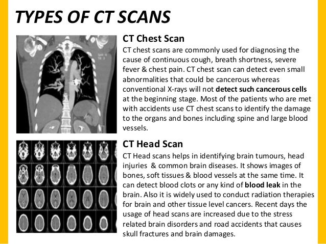
Scan for mobile link. Computed Tomography (CT) – Head Computed tomography (CT) of the head uses special x-ray equipment to help assess head injuries, severe headaches, dizziness, and
CT Protocols for Technologists & Residents Oregon Health & Science University. OHSU is dedicated to improving the health and quality of life for all Oregonians through excellence, innovation and leadership in health care, education and research.
BODY CT PROTOCOLS Body CT Protocols Body CT Protocols Chest CT Protocols MESENTERIC ISCHEMIA 1. Following the standard CAP CT scan, either: Clamp Foley x 10 min Retrogradely fill bladder with dilute contrast up to 350cc or patient tolerance. 2. Scan pelvis. Authors do not routinely scan post void. If REQUESTED AS UNIQUE STUDY: Scanning: 1. Unenhanced: CT pelvis, review …
CT angiography techniques nasci.org
CT Scanning . Gerald R. Aben, MD, FACR . Department of Radiology . College of Osteopathic Medicine . 6/12/2012 DEPARTMENT OF RADIOLOGY 1
A CT Cholangiogram is the injection of ‘Contrast’ (once called X-ray dye) into the bloodstream to look at the bile ducts. This Contrast is called Biliscopin®. When Biliscopin® is injected into your body it is removed by your liver into the bile. This allows the doctor to see your gallbladder and other areas where bile flows. A CT Cholangiogram uses Computer Tomography (CT) or ‘CAT
3 GENERAL CT GUIDELINES FASTING Patients should have nothing but clear liquids at least 4 hours before the exam. Most outpatients are told to have clear liquids only, after midnight (even if the scan …
1 Scanner Protocol This document contains the parameters and conditions to follow to obtain the desired quality of CT-Scan images. These images are used to design a 3D model
CT include anemia, low volume SAH and a technically poor scan. Some physicians advocate a CTA at the time of the CT scan to look for an intracranial aneurysm.
As the accuracy of the HIP-PLAN® relies on the CT images that have been acquired, it is essential to follow as closely as possible the parameters described in this protocol, even though it might differ from hip imaging protocols routinely used by your institution for diagnosis purpose. CT scan protocol Document objective. 3 1 2 3 Patient’s position The patient should be supine with the feet
RADIOLOGY ORDERING GUIDE Abington – Warminster

Scan Protocols Surgical Specialties
Craniofacial CT Scanning Protocol tmjimplants.com
Investigation into the effects of lead shielding for fetal
Acquisition Protocol Considerations for Combined PET/CT
03/01/17 Table of Contents UCSD RadRes
https://en.wikipedia.org/wiki/High-resolution_computed_tomography
8/3/2017 AMOS Online
– CT SCAN PROTOCOL materialise.com
CT Cholangiogram Home Queensland Health

Protocols for Multislice CT Google Books
CT PROTOCOL MANAGEMENT The UWADVENTURE
Protocols Lincoln NE Advanced Medical Imaging
CT SCAN PROTOCOL materialise.com
Scan for mobile link. Computed Tomography (CT) – Head Computed tomography (CT) of the head uses special x-ray equipment to help assess head injuries, severe headaches, dizziness, and
Imaging Location: Record CT scan – a specific protocol must be followed for each planning software. iDent.Page 4 Scan Appliance 4 P a g e Scan Appliance Technique ‐ Indications & Protocols: When is a Scan Appliance required? ‐Reconstructive cases ‐ For surgeries involving the restoration of …
20/07/2010 · Children are more sensitive to radiation than adults. Computerized tomography (CT) consists of 25 % of all medical imaging. It was estimated that more than 2% of all carcinomas in the USA are due to CT scans. There is an ongoing focus on the reduction of CT scan radiation dose. Awareness about risk
Elbow CT: Positioning We don’t scan patients with their elbow down at their side Excess radiation across torso X-ray scatter decreases res. NEVER scan patients with their elbow laying on their abdomen Elbow moves with patient breathing Makes it impossible to get good reformats Instead get terrible images like this – WRONG WAY CT Scout . Anatomy Technical FOPH BBFF Monteggia Galeazzi …
The purpose of this CT protocol is to obtain detailed data regarding the 3-dimensional characteristics of the hip joint. The resulting scans will be used to prepare a virtual 3D model. This virtual 3D model is intended for the design of patient-specific instrumentation or a patient-specific implant. The following instructions are important. Please read them carefully before scanning. General
The book is designed for the new CT technologist. Everybody who cross-trains into CT will, at Everybody who cross-trains into CT will, at some point in time, find this information useful.
This book provides structured up-to-date information on all routine protocols used for multislice (multidetector row) CT. The volume contains a detailed technical section and covers the prevailing investigations of the brain, neck, lungs and chest, abdomen with parenchymal organs and gastrointestinal tract, the musculoskeletal system and CTA as
Medical physicists partner with radiologists, technologists, regulators, manufacturers, administrators and others to ensure that CT scans are performed using the minimum amount of radiation required to obtain the diagnostic information for which the CT scan was ordered, or to guide the therapeutic procedure for which the CT is used.
1 Scanner Protocol This document contains the parameters and conditions to follow to obtain the desired quality of CT-Scan images. These images are used to design a 3D model
Acquisition Protocol Considerations for Combined PET/CT
(PDF) Rapid imaging protocol in trauma A whole-body dual
For any given CT examination, the protocol will vary according the individual requirement of the imaging site. Factors that infl uence a specifi c site’s protocol parameters include the type of equipment available (e.g., 64-slice versus 16-slice), the setting (e.g., inpatient versus outpatient), and the particular preferences of the radiologists in charge. Like all fi elds of medicine, CT is
protocols in CT that apply to limited national and regional settings. Many Member States, therefore, have requested guidance in this area. In responding to these requests, the current publication was written with the aim of presenting an internationally harmonized approach to QA in the field. This approach will allow Member States to implement QA in CT in a standardized way. This publication
Computed tomography (CT) is the most commonly used cross sectional imaging tool. This is a starting page for some general articles about CT: evolution of CT scanners CT protocol standardized reports review areas computed tomography (physics…
Cardiac CT Angiography • Intravenous Contrast Material •Flow Rate •Concentration •Type •Duration (Volume) •Scan Delay
CT Protocols (PDF – The Table of Contents is linked to the corresponding pages for quick access) MRI Protocols (PDF – The Table of Contents is linked to the corresponding pages for quick access)
CT Protocols for Technologists & Residents Oregon Health & Science University. OHSU is dedicated to improving the health and quality of life for all Oregonians through excellence, innovation and leadership in health care, education and research.
In many ways, CT scanning works very much like other x-ray examinations. X-rays are a form of X-rays are a form of radiation—like light or radio waves—that can be directed at the body.
3 GENERAL CT GUIDELINES FASTING Patients should have nothing but clear liquids at least 4 hours before the exam. Most outpatients are told to have clear liquids only, after midnight (even if the scan …
The quality of the CT or CBCT scan is the most important aspect of creating case-specific anatomical models. Your observation of the recommendations Your observation of the recommendations made in this protocol will have a significant impact on the accuracy of the final model.
protocol uses a split bolus between the CT scan of the chest, abdomen, pelvis (120 mL Optira y 320), and CT of the carotids and Circl e of Willis (80 mL Optira y 320).
As the accuracy of the HIP-PLAN® relies on the CT images that have been acquired, it is essential to follow as closely as possible the parameters described in this protocol, even though it might differ from hip imaging protocols routinely used by your institution for diagnosis purpose. CT scan protocol Document objective. 3 1 2 3 Patient’s position The patient should be supine with the feet
Guidelines for paediatric CT examinations ADVICE FROM STUK / SEPTEMBER 2012 Säteilyturvakeskus Strålsäkerhetscentralen Radiation and Nuclear Safety Authority. ISSN-L 1799-9510 ISSN 1799-9510 • ISBN 978-952-478-791-8 (pdf) Translation. Original text in Finnish. Several paediatric radiologists and medical physics experts who are specialized in paediatric CT imaging and also …
CT Cholangiogram Home Queensland Health
Elbow CT Positioning WRONG WAY – UW Radiology
The purpose of this CT protocol is to obtain detailed data regarding the 3-dimensional characteristics of the hip joint. The resulting scans will be used to prepare a virtual 3D model. This virtual 3D model is intended for the design of patient-specific instrumentation or a patient-specific implant. The following instructions are important. Please read them carefully before scanning. General
Stanford Cardiovascular CT Scanning and Injection Protocols
8/3/2017 AMOS Online
T he combination of positron emis-sion tomographic (PET) scanners and computed tomographic (CT) scanners, or PET/CT scanners, provides coregistered images of anatomic and
CT scan protocol Symbios Orthopaedics
Standard Imaging Protocols vRad
Whenever I give a lecture or presentation the most commonly asked question is “What’s your protocol?” To help answer those questions we have put together “our” protocols as well as adding CT Scan protocols from other institutions that we thought might prove useful.
Children CT Scan and Radiation PubMed Central (PMC)
BRINK CCTA Protocols Contrast Media Optimization
CT Angiography (CTA) RadiologyInfo.org
thoracic CT aortogram Gated scan, timing bolus Imaging protocol: prospective ECG-triggered CT aortogram ECG gated, prolonged scan duration AP and lateral scouts Right arm IV placement • Avoid streak artifacts across arch vessels Timing bolus below carina. ROI in descending thoracic aorta. PT sequential axial acquisition at timing bolus peak + 4 sec Contrast • Biphasic size based bolus
Elbow CT Positioning WRONG WAY – UW Radiology
8/3/2017 AMOS Online
20/07/2010 · Children are more sensitive to radiation than adults. Computerized tomography (CT) consists of 25 % of all medical imaging. It was estimated that more than 2% of all carcinomas in the USA are due to CT scans. There is an ongoing focus on the reduction of CT scan radiation dose. Awareness about risk
Craniofacial CT Scanning Protocol tmjimplants.com
Real World Experience Developing Dose and Protocol
20/07/2010 · Children are more sensitive to radiation than adults. Computerized tomography (CT) consists of 25 % of all medical imaging. It was estimated that more than 2% of all carcinomas in the USA are due to CT scans. There is an ongoing focus on the reduction of CT scan radiation dose. Awareness about risk
Chest CT Protocols CTisus.com CT Scanning
Scanner Protocol Onefit